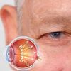
What is Eye Pressure (Glaucoma)? How to Treat?
Ocular hypertension is a chronic disease in which the intraocular pressure (IOP) goes out of balance and puts constant pressure on the optic nerve. It progresses silently at first, but if not diagnosed early, it can lead to permanent blindness. When the fluid constantly produced within the eye is not sufficiently filtered through the trabecular meshwork, the internal pressure rises slowly; This rise narrows the field of vision, especially starting from the outer aspects. Those with a family history of glaucoma, those over the age of fifty, those with diseases that affect vascular health such as diabetes-hypertension, and those who use cortisone for a long time are in the risk group for this insidious condition, which can be detected at an early stage even with a simple tonometer measurement.
What is Eye Pressure (Glaucoma)?
Eye pressure, medically known as glaucoma, describes the increase in pressure that occurs when the drainage of the aqueous humor fluid, which is constantly produced and circulating within the eye, slows down. Normally, intraocular pressure remains in the range of 10–21 mmHg; Microscopic channels called trabecular meshwork transfer this fluid into the bloodstream. When the channels are blocked or fluid production is excessive, the pressure increases, the eyeball stretches like a "balloon" and puts constant pressure on the optic nerve. This pressure reduces the blood flow of the capillaries feeding the nerve fibers; Fibers that cannot be nourished are irreversibly damaged. The most insidious aspect of glaucoma is that it narrows the peripheral visual field without causing any symptoms in the early stages. The patient usually does not realize it until central acuity is affected; At this stage, most of the nerve damage is permanent.
What Causes Eye Pressure (Glaucoma)?
Ocular hypertension occurs when the delicate balance between the production and drainage of intraocular fluid (aqueous humor) is disrupted. Normally, fluid is produced in the ciliary body and passes into the bloodstream through microscopic channels called trabecular meshwork, keeping the intraocular pressure within the range of 10–21 mmHg. If these channels become blocked, narrowed, or fluid production increases, the pressure rises; The increased pressure puts pressure on the optic nerve and causes irreversible fiber loss over time.
- Genetic Predisposition — In individuals with a family history of glaucoma, the drainage angle may be congenitally narrower or the network structure may be more impermeable.
- Ocular Trauma — Impacts to the eye disrupt the network structure, paving the way for the pressure to rise silently.
- Long-Term Cortisone — Chronic use of steroid drops, tablets or inhalers can cause protein accumulation in the channels and increased resistance.
- Systemic Diseases — Diabetes, hypertension, sleep apnea and thyroid disorders reduce intraocular blood flow, making nerve fibers more sensitive to pressure.
- Eye Structural Features — In high myopia or hyperopia, anatomical differences of the eyeball may lead to narrowing of the drainage angle.
- Sudden Iris Block — In closed-angle glaucoma, the iris root blocks the drainage angle like a "plug"; fluid cannot come out and the pressure skyrockets within hours, creating severe pain, nausea and rapid vision loss.
Types of Glaucoma
- Open Angle Glaucoma — It moves slowly; narrowing of the trabecular meshwork causes pressure to rise silently.
- Closed Angle Glaucoma — The iris root abruptly closes the drainage angle; Severe pain, nausea and blurred vision occur, urgent treatment is required.
- Normal Tension Glaucoma — Even if the pressure remains within the normal range, the optic nerve is damaged due to low blood flow; Blood pressure measurement alone does not provide sufficient diagnosis.
- Congenital Glaucoma — Drainage channels are not developed at birth; Inability to look at the light, watery eyes and large cornea are noted in the baby.
- Secondary Glaucoma — Uveitis develops after diseases such as eye trauma, long-term steroid use or diabetes.
Risk Factors
- Family History — Having a family history of glaucoma increases the risk.
- Advanced Age — Being fifty years old and above is in the risk group.
- Refractive Defects — High myopia or hyperopia carries a risk.
- Systemic Diseases — Diabetes, hypertension, and sleep apnea syndrome increase the risk.
- Cortisone Use — Long-term cortisone treatment poses risks.
- Eye Trauma — Having eye trauma puts you in the risk group.
Symptoms of Eye Pressure
- Visual Field Narrowing — Noticing gradual narrowing of the outer edges of the visual field.
- Rings of Light — Seeing colored rings of light in the dark or when looking into bright headlights.
- Headache and Eye Pain — Feeling dull pressure around the eyes and a mild headache when you wake up in the morning.
- Reading Difficulty — Skipping lines or noticing objects coming from the side late while reading.
- Closed Angle Attack — Severe eye and forehead pain, nausea, blurred vision in a sudden closed angle attack.
- Loss of Night Vision — Weakened perception of stairs or steps in dim light; Excessive glare from oncoming headlights while driving at night.
- Tunnel Vision — In the advanced stage, although the central vision remains clear, the side vision remains in a "tunnel" form.
Eye Pressure Diagnosis
Diagnosis of eye pressure is not just a simple pressure measurement with a tonometer; Even if the pressure value remains within normal limits, damage to the optic nerve may begin. Therefore, diagnosis consists of five basic stages that complement each other: intraocular pressure measurement with tonometer, drainage angle evaluation with gonioscopy, optic nerve head examination with biomicroscope and OCT, visual field test and pachymetry measuring corneal thickness. When all these data are brought together, a diagnosis of "glaucoma risk", "early glaucoma" or "advanced glaucoma" is made and a personalized treatment schedule is created.
Eye Pressure Treatment
Treatment of eye pressure basically aims to protect the optic nerve by reducing the intraocular pressure to the target range, and in most patients, the first step is drop treatment. Prostaglandin analogues widen drainage channels, while beta blockers and carbonic anhydrase inhibitors reduce fluid production. Dropping the drug at the same time every day slows down the loss of the optic nerve as it prevents pressure fluctuations. When sufficient reduction cannot be achieved with treatment medications, laser methods such as selective laser trabeculoplasty or peripheral iridotomy are used. If the pressure remains high, surgical options such as trabeculectomy or micro stent implants are used. Treatment is not limited to medical interventions only; Regular exercise, smoking and caffeine restriction, salt reduction and a diet rich in omega-3 also support nerve nutrition.
Lifestyle and Preventive Recommendations
- Exercise — Moderately brisk walking five days a week improves circulation.
- Cigarettes and Caffeine — Avoiding smoking and excessive caffeine protects vascular health.
- Head Position — Keeping the head slightly above heart level during screen breaks limits pressure fluctuation.
- Salt Restriction — Restricting salt consumption improves nerve nutrition by balancing systemic blood pressure.
- Polarized Glasses — Using polarized glasses that reduce light reflections increases visual comfort.
Frequently Asked Questions
Even if the pressure decreases to the target, the drops last a lifetime to protect the optic nerve. Discontinuing the medication may cause the pressure to silently rise again and cause damage to progress.
Yes, but wait at least five minutes between each drop. Thus, the first drop is absorbed without washing and the second drop remains in the eye at the effective dose.
In most patients, the number of drops decreases, but it is rarely possible to stop completely. In the controls after the laser procedure, the drop plan is rearranged according to the pressure course.
Trabeculectomy or microstenting can permanently reduce pressure, but scar tissue formation can narrow the channel. Early intervention is performed with regular check-ups and supportive drops are added if necessary.
Moderate walking and swimming help balance the pressure. Avoid holding your breath and upside down yoga poses while lifting weights; these may cause temporary pressure spikes.
No. No herbal product can replace drops, laser or surgery. Unless your doctor approves, using herbal supplements may disrupt the treatment.
Common treatments for glaucoma are drops, laser and surgery. Intraocular injection (anti-VEGF) is often used for different diseases such as diabetic retinopathy; It is not routine in glaucoma.
Soft lenses may reduce the absorption of drops. Hard lenses may irritate the filter under the lid after surgery. It is recommended that you stop using contact lenses or switch to prescription glasses by consulting your doctor.
Drops begin to reduce pressure from the first day; however, visual field and OCT results must remain stable over several checks to confirm that nerve damage has stopped.
Early surgery is a priority in congenital glaucoma; Drops and laser involve different doses and techniques than the adult plan. Pediatric patients should be monitored in centers with experience in pediatric eye surgery.