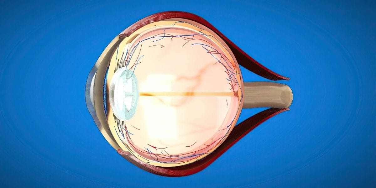
High eye pressure — medically known as glaucoma — is one of the leading causes of permanent blindness worldwide, yet in many patients it progresses without any symptoms. Also known as the "silent thief of sight," by the time symptoms are noticed, permanent damage has often already occurred. In this article, we comprehensively cover the symptoms of eye pressure, risk factors, and why early diagnosis is so critical.
What Is Eye Pressure (Glaucoma)?
Eye pressure occurs when intraocular pressure (IOP) rises above normal levels. When the fluid called "aqueous humor," which is continuously produced and renewed inside the eye, cannot drain sufficiently through the drainage channels, pressure builds up. Over time, this elevated pressure damages the optic nerve, leading to visual field loss and, in advanced cases, blindness.
Normal intraocular pressure is between 10 and 21 mmHg. Values above 21 mmHg are defined as ocular hypertension, though it should be noted that glaucoma can develop even below 21 mmHg in some patients (normal-tension glaucoma).
Types of Glaucoma
- Primary open-angle glaucoma: The most common type. The drainage channels are anatomically open but have lost their function. It can progress for years without symptoms.
- Acute angle-closure glaucoma: The drainage canal closes suddenly; it manifests with severe eye pain, headache, nausea, and blurred vision. Requires emergency intervention.
- Normal-tension glaucoma: Optic nerve damage develops despite intraocular pressure being within normal limits. Diagnosis is challenging.
- Secondary glaucoma: Develops due to another eye disease or medication use (e.g., long-term corticosteroid use).
- Congenital glaucoma: Seen at birth or in early childhood; may present with abnormal pupil size, sensitivity to light, and excessive tearing.
Eye Pressure Symptoms: Why Is It So Hard to Notice?
The most dangerous feature of glaucoma is that its primary open-angle form — the most common type — causes no symptoms for many years. Visual field loss starts from the periphery; since the brain compensates for this loss, the patient usually does not notice it. By the time damage reaches central vision, significant vision loss may have already occurred.
Symptoms in Acute Angle-Closure Glaucoma (Emergency)
Although rare, the symptoms of this type are much more pronounced:
- Sudden onset of severe eye pain
- Headache (especially in the forehead area)
- Nausea and vomiting
- Blurred vision
- Colored halos around light sources
- Redness in the eye
When these symptoms appear, you should visit an emergency eye unit without delay.
Possible Symptoms in Chronic (Open-Angle) Glaucoma
In chronic glaucoma, symptoms may be very subtle or completely absent. In advanced stages of the disease, the following changes may be noticed:
- Narrowing or blurring at the edges of the visual field (peripheral vision)
- Difficulty seeing in the dark
- Discomfort in bright light
- Sensation of certain objects disappearing and reappearing
- Unexplained changes in eyeglass prescription
These findings usually appear when a significant portion of the visual field has already been lost.
Eye Pressure Risk Factors
Even without any symptoms, individuals with the following risk factors should prioritize regular eye examinations:
| Risk Factor | Description |
|---|---|
| Advanced age | Risk increases significantly in individuals over 60 years of age |
| Family history | Risk increases 4-9 times if a parent or sibling has glaucoma |
| High myopia | A longer axial length makes the optic nerve more vulnerable |
| High intraocular pressure | Values above 21 mmHg require close follow-up |
| Diabetes and hypertension | Vascular risk factors increase the risk of glaucoma |
| Long-term corticosteroids | Systemic and inhaled use, including eye drops |
| History of eye trauma | May damage the drainage canal |
| Thin cornea | May cause true IOP to appear lower than it actually is |
How Is Eye Pressure Measured and Diagnosed?
Intraocular pressure measurement is an integral part of a standard eye examination. However, a glaucoma diagnosis does not rely solely on pressure measurement; it requires multiple assessments:
Diagnostic Methods
- IOP measurement with tonometry: Goldmann applanation tonometry is the gold standard method. Air-puff (non-contact) tonometers are less precise.
- Gonioscopy: An examination performed with a mirrored lens to determine the type of drainage angle (open/closed).
- Optic disc examination: Evaluates the "cup-to-disc" ratio and blood supply at the optic nerve head.
- Visual field test (perimetry): Maps the central and peripheral visual field and shows the localization of vision loss.
- OCT (Optical Coherence Tomography): Measures retinal nerve fiber layer thickness at the micron level and can detect structural damage before visual field loss begins.
- Pachymetry: Measures corneal thickness to adjust tonometry readings.
Eye Pressure (Glaucoma) Treatment
Damage in glaucoma is irreversible; therefore, the goal of treatment is to preserve existing vision and halt the progression of the disease.
Medication Therapy
The first choice is usually eye drop therapy that lowers intraocular pressure. Prostaglandin analogs, beta-blockers, carbonic anhydrase inhibitors, and alpha-2 agonists lower intraocular pressure through different mechanisms. Treatment effectiveness is evaluated through regular follow-up examinations.
Laser Treatments
Selective laser trabeculoplasty (SLT) stimulates the trabecular meshwork to increase drainage and is used as an alternative or complement to drop therapy in certain patient groups.
Surgical Treatments
When medication and laser therapy do not provide an adequate response, trabeculectomy (filtration surgery), tube implants, or minimally invasive glaucoma surgery (MIGS) may be performed.
Birinci Göz Hospital Approach to Early Glaucoma Diagnosis
At Birinci Göz Hospital, a comprehensive evaluation protocol is applied to patients suspected of having glaucoma. Visual field analysis, retinal nerve fiber layer measurement with OCT, and gonioscopy findings are evaluated together to create a personalized follow-up and treatment plan. In particular, individuals over the age of 40 with a first-degree relative diagnosed with glaucoma are advised to have an eye pressure check at least once every two years.
Conclusion
Glaucoma is a serious disease that can be successfully controlled when diagnosed early, but can lead to permanent vision loss if neglected. It is vital to know that the thought "I have no symptoms, so there is no problem" poses a life-altering risk in this disease. If you have risk factors or are over 40, do not postpone your regular eye examination; your eyes may be silently giving you a sign.
Frequently Asked Questions
Does high eye pressure always mean glaucoma?
No. Individuals with intraocular pressure above 21 mmHg but without optic nerve damage are diagnosed with "ocular hypertension." These individuals require close follow-up, and the decision to treat is made based on their risk profile.
Can eye pressure drop on its own?
Generally, no. Although pressure may fluctuate from time to time in individuals diagnosed with ocular hypertension, normalization without treatment is not expected.
Do children of someone with glaucoma inherit the disease?
Having a first-degree relative with glaucoma increases the risk by 4 to 9 times. Therefore, individuals with a family history of glaucoma are advised to undergo a dilated eye exam at least every two years starting at age 40.
Does eyeglass prescription change if eye pressure is high?
Elevated intraocular pressure does not directly change refractive error. However, visual field narrowing as glaucoma progresses can have a serious impact on reading and functional vision.
Does glaucoma treatment last a lifetime?
Primary open-angle glaucoma is a chronic disease, and its treatment is usually lifelong. Laser therapy may reduce the need for drops in some patients, but it may not eliminate it completely.