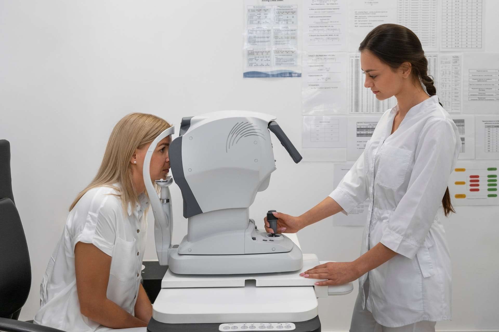
OCT (Optical Coherence Tomography) is a high-tech imaging method that examines the retina of the eye at the micron level. Thanks to OCT (Macula), even the smallest changes in the macular layer can be detected and early diagnosis can be made.
Why is the Macula Important?
The macula is located in the middle of the retina layer and provides central vision of the eye. Defects in the macula can cause decreased visual acuity, changes in color perception, and curvature or darkening of vision.
Why is OCT (Macula) Done?
- Complaints of blurring, curvature or darkening of vision
- Suspicion of macular degeneration
- Monitoring diabetic retinopathy or retinal edema
- Retinal vascular occlusions
- Evaluations before and after retinal surgery
- Assessment of the risk of age-related vision loss
How is OCT Procedure Done?
OCT is a completely painless, fast and contactless method. The pupil is usually not dilated during the procedure. The patient sits in front of the device and looks at a fixed point; The device takes high-resolution cross-sectional images of the retina layers within seconds. Both eyes can be examined separately.
Which Diseases Are Tracked with OCT?
- Age-related macular degeneration
- Diabetic retinopathy and diabetic macular edema
- Retinal vascular occlusions
- Macular hole and cystoid edema
- Recovery follow-up after retinal surgery
Frequently Asked Questions
Is OCT testing painful?
No. OCT is a completely painless, non-contact and rapid test. After the procedure, the patient can return to daily activities immediately.
Can the pupil be dilated for OCT?
Usually not necessary. In some cases, the doctor may recommend drops for a clearer image; In this case, temporary slight blurriness may occur.
Who is OCT testing recommended for?
Regular check-ups are of great importance, especially for people over the age of 50, diabetics and anyone with vision complaints.